Sourcing is the question that separates a specification from a story. Two vials labelled the same way can come from different tissues, be enriched by different methods, and carry different proportions of the cells the label names. For a physician or laboratory evaluating cellular material, where it came from and how it was separated out is the part that determines what the rest of the paperwork is worth.
This page covers the tissues Muse cells have been isolated from, the two methods used to harvest them, and why the umbilical cord has become the reference source. For the underlying biology, start with
what Muse cells are and how they were identified.
Muse cells have no dedicated source tissue
Muse cells are not sourced from a dedicated tissue. They are a rare subpopulation resident within mesenchymal tissue, so the answer to where Muse cells come from is: wherever mesenchymal cells come from, in small and variable proportions.
They were first reported from adult human bone marrow by Kuroda and colleagues in PNAS in 2010 and have since been identified across a range of tissues. That distributed origin is the reason sourcing matters commercially: the tissue determines the yield, the donor profile, and the consistency, and none of those are visible from the product name.
The tissues Muse cells have been isolated from
Published work has recovered SSEA-3-positive Muse cells from bone marrow, adipose tissue, dermal fibroblasts, peripheral blood, amniotic membrane, and umbilical cord, as reviewed by Gu and colleagues in Frontiers in Cell and Developmental Biology in 2024.
Bone marrow and umbilical cord are the two sources that dominate the literature. They differ in almost every way that matters operationally. Bone marrow requires an aspirate from a living adult donor, with the donor burden and consent complexity that it implies. The umbilical cord is recovered after birth.
The yield figures need to be read carefully, because two different denominators circulate. In fresh bone marrow, Muse cells are on the order of 0.01–0.03% of mononucleated cells, roughly one cell in three thousand. In cultured mesenchymal populations, the fraction runs between 1% and 6% depending on source and handling. Those are not the same measurement, and comparing one against the other overstates the difference between tissues. On a like-for-like basis, cultured mesenchymal cells, Kushida and colleagues describe the umbilical cord fraction as comparable to or higher than that found in bone marrow, adipose, and dermal sources.
Donor age is the variable underneath both. Adult sources carry whatever the donor’s tissue carried, age, exposure history, replicative history, and a bone-marrow aspirate is, by definition, from an adult. Perinatal tissue does not. That is a difference in starting material rather than a claim about performance, and it is one of the reasons the two sources are rarely interchangeable in a process that has been validated on one of them.
How Muse cells are harvested
Two methods are in use, and they are not interchangeable. Anyone asking how Muse stem cells are harvested should expect a supplier to name which one was used.
The first is stress selection. The original 2010 work applied prolonged trypsin incubation to mesenchymal cultures, sustained proteolytic stress under which most of the population died, and a surviving fraction proved to carry pluripotency markers and triploblastic capacity. Later work has reproduced the same selection with long collagenase exposure, serum deprivation, hypoxia, and low temperature. The method selects for survival.
The second is marker sorting. Tissue is cultured to a mesenchymal population, then SSEA-3-positive cells, often SSEA-3 and CD105 double-positive, are separated by fluorescence-activated or magnetically-activated cell sorting. The method selects based on marker expression at the moment of sorting.
There is a second reason the method matters, and it is about consistency rather than identity. Stress selection is a biological filter, and its output depends on how the starting population responded to the insult on that particular day, with that particular donor’s cells. Marker sorting is an instrument-defined cut against a gate someone set. Neither is inherently better, but they fail differently, and the failure mode is what a laboratory needs to know when a lot behaves unlike the one before it. A supplier who can state the method, the gate, and the operator’s acceptance criteria is describing a process under control.
Wharton’s jelly and the UC-MSC question
Two questions come up constantly here, and they are really the same question asked at different resolutions: are Muse cells the same thing as UC-MSC, and do they come from the umbilical cord generally or from Wharton’s jelly specifically?
The clearest answer in the literature comes from
Kushida and colleagues in Cellular and Molecular Life Sciences in 2024, from Mari Dezawa’s group. Working with human umbilical cord, they located SSEA-3-positive cells sparsely distributed as individual cells, 9.5 to 13.8 micrometres across, within Wharton’s jelly the loose connective tissue of the cord. Cord tissue was minced and cultured to a mesenchymal population, and the SSEA-3-positive fraction was then separated by sorting.
So both questions resolve cleanly. Muse cells are not an alternative to UC-MSC; they are a fraction within it. And within the cord itself, Wharton’s jelly is where they were found.
That paper also gives the fraction a number: SSEA-3-positive cells accounted for 5.2 ± 0.3% of umbilical cord mesenchymal cells comparable to or higher than the proportions reported from bone marrow, adipose tissue and dermal sources, which sit in the 1–6% range across cultured mesenchymal populations.
Why the umbilical cord has become the reference source
Three reasons, and only one of them is biological.
Yield: at around 5% of the mesenchymal population, the cord is at the top of the reported range rather than the bottom.
Access: as the same authors note, umbilical cord can be obtained as medical waste after birth, without donor burden and without the ethical complexity attached to other sources.
Cell character: single-cell RNA sequencing in that study placed the gene expression profile of cord-derived Muse cells closer to human post-implantation blastocysts than to Muse cells from adult tissue, with lower methylation across differentiation-related genes, and around 20% of them lacking the X-inactivation marker XIST a feature not seen in adult-tissue Muse cells. Whether that profile carries any functional advantage is an open research question, and this page does not claim one.
The phrase “Dezawa Muse cells”
MUSE cells are a distinct cell population studied in scientific research for their unique biological characteristics and potential applications in regenerative medicine research. Early research and characterization of MUSE cells involved Mari Dezawa and collaborators, whose published work contributed to the scientific foundation for subsequent MUSE cell studies.
It is important, however, to distinguish the scientific concept of MUSE cells from specific commercial products, companies, or trademarks.
CellGenic MUSE Cells are not Dezawa MuseCells® and are not affiliated with Muse Cell Innovations. CellGenic does not use “Dezawa Muse Cells” as a product name or branding designation. References to Dezawa or other researchers are included only to provide accurate historical and scientific context.
For researchers and physicians evaluating MUSE cells, important considerations include cell sourcing, isolation methods, identity, purity, characterization, manufacturing processes, and quality controls.
CellGenic MUSE Cells are independently developed and positioned as a research product, supported by CellGenic’s own manufacturing and quality-control platform. Where supported by appropriate product documentation, specifications such as greater than 90% MUSE cell purity can be considered when evaluating product quality.
This distinction allows researchers to explore MUSE cell research while understanding that CellGenic’s product is independently positioned and should not be confused with third-party branded products or organizations.
Where CellGenic’s material comes from
Four facts, and deliberately only four. Everything else about a given lot belongs on that lot’s paperwork rather than on a page like this one.
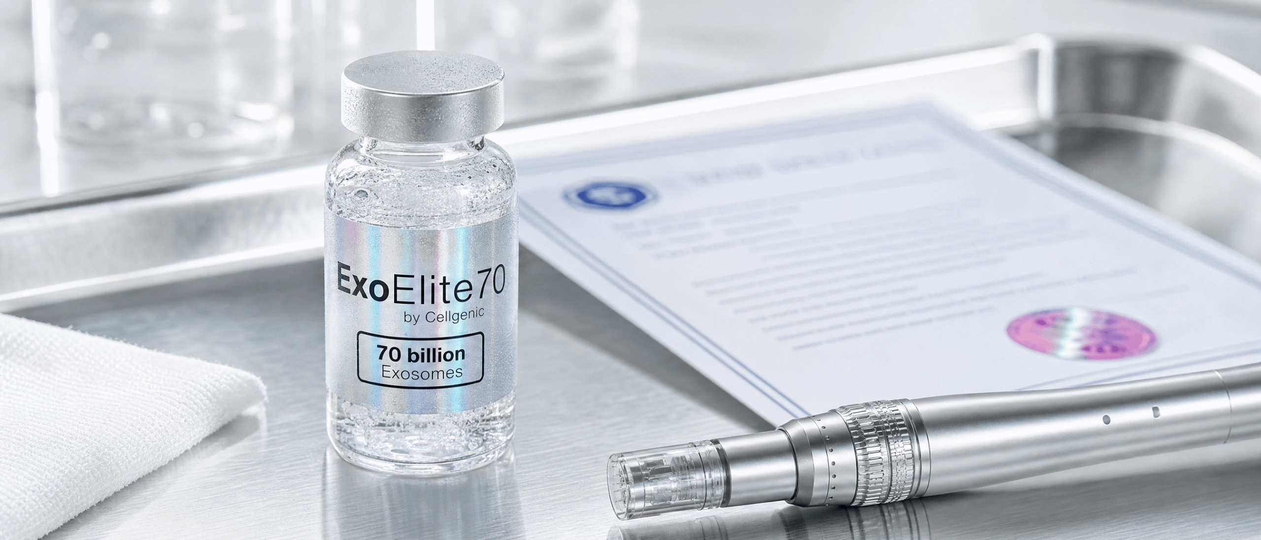
Manufacturing is cGMP. Processing is performed in an ISO 7 classified cleanroom. A certificate of analysis is issued for every batch, identified by batch number. And access to that documentation, and to ordering, requires provider verification.
That last one is not a formality. Batch documentation is released to verified accounts because the material is supplied to licensed physicians, clinic operators and researchers, and the verification step is what establishes that.
What to establish before you order
Sourcing questions are answerable in specifics or they are not answerable at all.
Which tissue the population was sourced from. Whether the Muse fraction was established by stress selection or by marker sorting. What percentage of the preparation was SSEA-3-positive, and whether that was measured on this batch or on a validation lot. Whether the certificate of analysis for the batch in hand can be read before ordering rather than after. And, because batch number is the identifier that ties all of it together, whether the batch number on the vial matches the one on the certificate.
Nothing on this page establishes that Muse cells treat, cure, prevent or diagnose any condition, and nothing on it supports use outside a properly authorised research setting under applicable regulation. Sourcing and characterisation are questions about material, not about clinical effect.
Common Questions
References
Sources cited or paraphrased in this article are listed alphabetically by first author.
REGULATORY DISCLAIMER · CELLS
These products are intended for laboratory research use only. They are not drugs, foods, cosmetics, or medical treatments and must not be used for any form of human or animal administration. All information provided is for educational and scientific reference only and the products should be handled exclusively by licensed, qualified professionals. Misbranding, misuse, or mislabeling of these products as therapeutic or consumable substances is strictly prohibited by law.

 Medically Reviewed By
— Written By
— Updated on September 16, 2026