
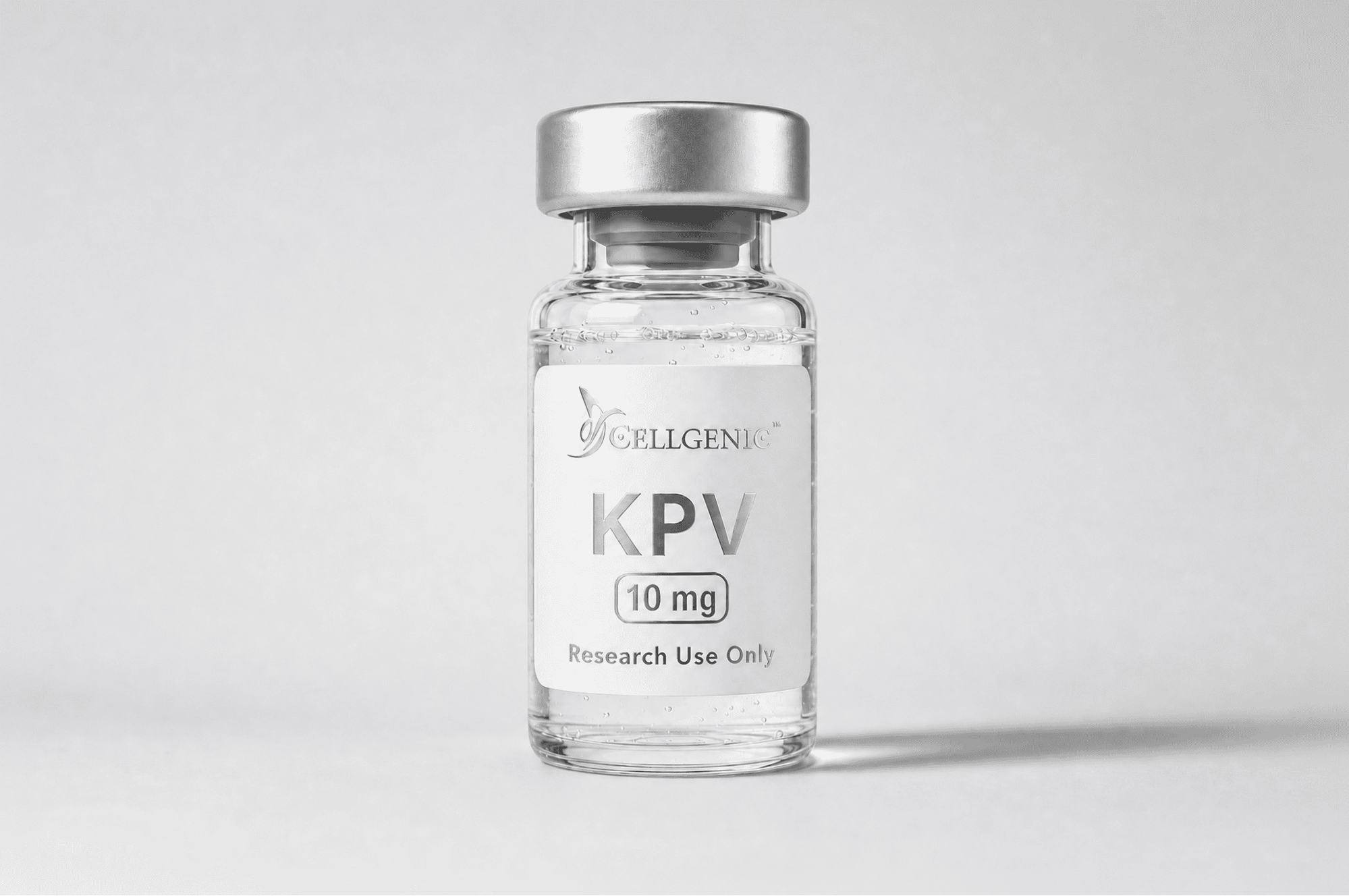
FOR RESEARCH USE ONLY. This article is for educational and informational purposes only, and is based on published scientific literature. It does not constitute guidance on human or animal use and should not be interpreted as such.
The KPV peptide is a tripeptide corresponding to residues 11–13 of alpha-melanocyte-stimulating hormone (alpha-MSH), consisting of three amino acids: lysine, proline, and valine. Published research has shown that KPV retains the parent hormone’s anti-inflammatory signaling activity without its melanogenic effects (2).
Studies have demonstrated that KPV modulates NF-κB signaling, the master transcription factor regulating inflammatory gene expression, without stimulating melanogenesis, a side effect associated with full-length alpha-MSH (2). This selectivity has made KPV a subject of published research across intestinal, dermal, and neurological tissue models (1).
This article examines the current state of KPV peptide research, covering its molecular origin, mechanism of action, key findings from published studies, and its role within CellGenic’s broader peptide catalog.
Key Takeaways
- KPV is a C-terminal tripeptide (Lys-Pro-Val) derived from alpha-MSH. Published research indicates it retains the parent hormone’s anti-inflammatory signaling activity without its melanogenic effects (2).
- The primary KPV mechanism of action, as described in published literature, involves inhibition of NF-κB activation by preventing IκBα degradation, reducing downstream production of pro-inflammatory cytokines including IL-1β, IL-6, and IL-12 (1)(2).
- In intestinal models, researchers found that KPV enters epithelial cells via the PepT1 transporter rather than melanocortin receptors, a unique uptake mechanism confirmed through PepT1-knockout studies (1).
- Published colitis research demonstrated significant reduction in disease severity with oral KPV administration in both DSS-induced and TNBS-induced models (1).
- CellGenic manufactures KPV in its cGMP facility with lot-traced Certificates of Analysis documenting purity, sterility, and mycoplasma testing for each production batch.
KPV Mechanism of Action: NF-κB Inhibition and Beyond
Published studies have characterized KPV’s primary anti-inflammatory mechanism as suppression of NF-κB (nuclear factor kappa-light-chain-enhancer of activated B cells), the master regulator of inflammatory gene expression across virtually all cell types (2).
NF-κB Pathway Inhibition
Under normal conditions, NF-κB is sequestered in the cytoplasm by its inhibitor protein IκBα. Inflammatory stimuli trigger IκBα phosphorylation and degradation, releasing NF-κB to translocate into the nucleus and activate pro-inflammatory gene transcription. Research has shown that KPV inhibits this cascade by preventing IκBα degradation, keeping NF-κB sequestered and reducing inflammatory mediator expression (1).
Published studies have documented dose-dependent reductions in NF-κB activity following KPV treatment, with corresponding decreases in downstream cytokine production including interleukin-1β, interleukin-6, interleukin-12, and interferon-gamma (2).
MAPK Pathway Modulation
Beyond NF-κB, recent research has demonstrated that KPV also modulates MAPK (mitogen-activated protein kinase) signaling. A 2025 study published in Tissue & Cell reported that KPV inhibited the ERK/p38 MAPK/NF-κB signaling axis in human keratinocytes exposed to fine particulate matter, reducing both inflammation and apoptosis through suppression of caspase-1 activation (3).
PepT1-Mediated Cellular Uptake
One of the most significant findings in KPV peptide research is its cellular entry mechanism. Rather than acting through melanocortin receptors, researchers found that KPV enters intestinal epithelial cells and colonic immune cells via the peptide transporter PepT1. This was confirmed in PepT1-knockout mouse studies, where KPV failed to produce any anti-inflammatory or anti-tumorigenic effects, demonstrating that PepT1-mediated uptake is essential for KPV’s biological activity in intestinal tissue (1).
KPV Research in Intestinal Inflammation
The most extensively published area of KPV peptide research is intestinal inflammation, where the PepT1-mediated uptake mechanism provides direct access to gut epithelial cells.
A landmark study published in Gastroenterology by Dalmasso et al. investigated KPV in two mouse models of colitis: DSS-induced and TNBS-induced. Oral administration of KPV significantly reduced disease severity in both models, with measurable decreases in myeloperoxidase activity and improved epithelial barrier function (1).
The study confirmed that KPV’s intestinal anti-inflammatory activity depends on PepT1-mediated intracellular uptake followed by direct NF-κB suppression, mechanistically distinct from classical melanocortin receptor signaling. Researchers investigating gut barrier pathways can explore CellGenic’s research peptides catalog for compounds relevant to intestinal inflammation models.
KPV Peptide and Dermatological Research
Keratinocyte Protection
A 2025 study published in Tissue & Cell demonstrated that KPV protected human keratinocytes from fine particulate matter (PM2.5)-induced damage by inhibiting the ERK/p38 MAPK/NF-κB signaling pathway and suppressing caspase-1-mediated apoptosis (3).
Wound Healing and Dermatitis Models
A published review of alpha-MSH tripeptides documented KPV’s effects across multiple dermatological models (2). In corneal wound studies, topical KPV application produced significantly smaller wound areas compared to controls after four days. In atopic dermatitis models using NC/Nga mice, researchers observed reduced clinical severity scores, decreased epidermal thickening, and lowered serum IgE levels. Both intravenous and topical KPV suppressed contact dermatitis reactions in published mouse studies (2).
KPV in Neurological Inflammation Research
A study published in PLoS ONE demonstrated that a single administration of KPV attenuated brain damage in a mouse model of traumatic brain injury through reduced neuroinflammation and decreased apoptosis (4). This finding, alongside the intestinal and dermatological data, indicates that KPV’s NF-κB inhibition has been observed across multiple tissue types in published research.
KPV Within CellGenic’s Multi-Peptide Research Platform
Based on published data, KPV’s NF-κB suppression via PepT1-mediated intracellular delivery is mechanistically distinct from other peptides in the CellGenic catalog (1), making it a complementary compound for multi-pathway research.
CellGenic’s KLOW Stack combines KPV with BPC-157 (nitric oxide modulation), TB-500 (actin regulation), and GHK-Cu (matrix remodeling). These four compounds have non-overlapping mechanisms for combinatorial research. Additional protocol bundles featuring KPV include the Chronic Immune Dysfunction & Aging bundle and the Neuro-inflammation & Neuropathic Pain bundle, while CellGenic’s peptide calculator supports reconstitution calculations for KPV at any vial size.
CellGenic’s KPV is available in 5 mg, 10 mg, and 50 mg lyophilized vials manufactured to ≥98% purity under cGMP conditions.
Why KPV Peptide Research Continues to Expand
Based on the published data reviewed above, KPV’s small molecular size, PepT1-mediated cellular uptake (1), and NF-κB inhibition (2) distinguish it from both full-length alpha-MSH and other anti-inflammatory peptides. Published literature now spans intestinal, dermal, and neurological tissue models, each confirming NF-κB inhibition as KPV’s consistent mechanism across tissue types.
For researchers investigating KPV peptide, CellGenic delivers cGMP-manufactured product with the purity documentation and batch traceability that controlled research demands.
FOR RESEARCH USE ONLY. This article is for educational and informational purposes only, and is based on published scientific literature. It does not constitute guidance on human or animal use and should not be interpreted as such.
Frequently Asked Questions
1. What is the relationship between KPV and alpha-MSH?
KPV is the C-terminal tripeptide (residues 11–13) of alpha-melanocyte-stimulating hormone (alpha-MSH). It consists of three amino acids (lysine, proline, and valine). Published research has shown that KPV retains alpha-MSH’s anti-inflammatory signaling activity without stimulating melanogenesis, making it a more selective research tool for studying NF-κB-mediated inflammatory pathways (2).
2. How does KPV enter cells to exert its anti-inflammatory effects?
In intestinal tissue, researchers found that KPV enters epithelial cells and colonic immune cells via the PepT1 peptide transporter rather than through melanocortin receptors (1). This was confirmed in PepT1-knockout mouse studies, where KPV failed to produce anti-inflammatory effects. Once inside the cell, published data show that KPV inhibits NF-κB activation by preventing IκBα degradation (1).
3. What tissue types has KPV been studied in?
Published KPV peptide research spans intestinal tissue (colitis models)(1), skin (keratinocyte protection, wound healing, atopic and contact dermatitis)(2)(3), and neurological tissue (traumatic brain injury models)(4). The common finding across all tissue types is NF-κB pathway inhibition and reduction of pro-inflammatory cytokine production.
4. What is the KLOW Stack and how does KPV fit into it?
CellGenic’s KLOW Stack is a four-peptide research bundle combining KPV with BPC-157, TB-500, and GHK-Cu. Each compound has been studied for its effects through a distinct molecular pathway. Published research has investigated KPV for NF-κB inhibition, BPC-157 for nitric oxide signaling, TB-500 for actin dynamics, and GHK-Cu for matrix remodeling. This allows researchers to design multi-pathway protocols with non-overlapping mechanisms.
5. How should KPV peptide be reconstituted for research use?
Add bacteriostatic water slowly along the vial wall, swirling gently. Do not shake. Use CellGenic’s online peptide calculator to determine the correct solvent volume for your target concentration. Store reconstituted KPV at 2–8°C and use within four to six weeks.
References
- Dalmasso G, Charrier-Hisamuddin L, Thu Nguyen HT, Yan Y, Sitaraman S, Merlin D. “PepT1-mediated tripeptide KPV uptake reduces intestinal inflammation.” Gastroenterology. 2008;134(1):166–178. PMC Full Text
- Brzoska T, Luger TA, Maaser C, Abels C, Böhm M. “α-Melanocyte-stimulating hormone and related tripeptides: biochemistry, antiinflammatory and protective effects in vitro and in vivo, and future perspectives for the treatment of immune-mediated inflammatory diseases.” Endocrine Reviews. 2008;29(5):581–602. PubMed
- Sung J, Ju SY, Park S, Jung WK, Je JY, Lee SJ. “Lysine-Proline-Valine peptide mitigates fine dust-induced keratinocyte apoptosis and inflammation by regulating oxidative stress and modulating the MAPK/NF-κB pathway.” Tissue & Cell. 2025;95:102837. ScienceDirect
- Bitto A, Polito F, Irrera N, et al. “Single administration of tripeptide α-MSH(11–13) attenuates brain damage by reduced inflammation and apoptosis after experimental traumatic brain injury in mice.” PLoS ONE. 2013;8(8):e71056. PMC Full Text