 Medically Reviewed By
— Written By
— Updated on September 16, 2026
On this page
Why Sourcing and Isolation Matter
- Source affects biology. Cells from different tissues show different growth rates, surface markers, and secreted products.
- Method affects purity. Two isolation techniques can produce preparations with very different particle counts and contaminant profiles.
- Documentation affects confidence. Without batch records, a buyer cannot judge consistency from one lot to the next.
Where Research Cells Come From
Common Tissue Sources
Mesenchymal stromal cells (MSCs) were first described in bone marrow, and they have since been isolated from several other tissues. Common sources include:
- Bone marrow: the most studied source historically
- Adipose (fat) tissue: easier to collect in larger volumes
- Umbilical cord tissue: including Wharton’s jelly, the gelatinous matrix around cord blood vessels
- Cord blood: a separate source with its own cell populations
What Are MUSE Cells?
Donor Consent and Screening
Responsible sourcing begins with the donor. A documented process generally includes:
- Informed consent for the collection and its intended use
- Screening for infectious disease markers
- Review of donor history and eligibility criteria
- Traceability that links each lot back to its raw material
These steps protect the people who donate and give downstream users a paper trail. Programs accredited to biobanking standards such as ISO 20387 follow written procedures for this kind of traceability.
How Cells Are Isolated and Expanded
Getting from raw tissue to a characterized cell product typically involves these stages:
- Tissue processing. The tissue is washed and either cut into small pieces (explant method) or broken down with an enzyme such as collagenase (enzymatic digestion).
- Plating and selection. Cells attach to plastic culture surfaces, and non-adherent material is washed away.
- Expansion. Cells grow through several passages under controlled conditions.
- Characterization. Cells are tested against defined criteria.
- Cryopreservation. Cells are frozen in controlled conditions for storage and transport.
Explant vs. Enzymatic Methods
How Cells Are Characterized
The International Society for Cellular Therapy proposed minimal criteria for defining MSCs in 2006 [3]. These include:
- Adherence to plastic under standard culture conditions
- Expression of specific surface markers (such as CD73, CD90, and CD105) and absence of others (such as CD45 and CD34)
- Ability to differentiate into bone, fat, and cartilage cells in vitro
Passing these criteria confirms identity. It does not show that a product works for any health purpose.
Where Exosomes Come From
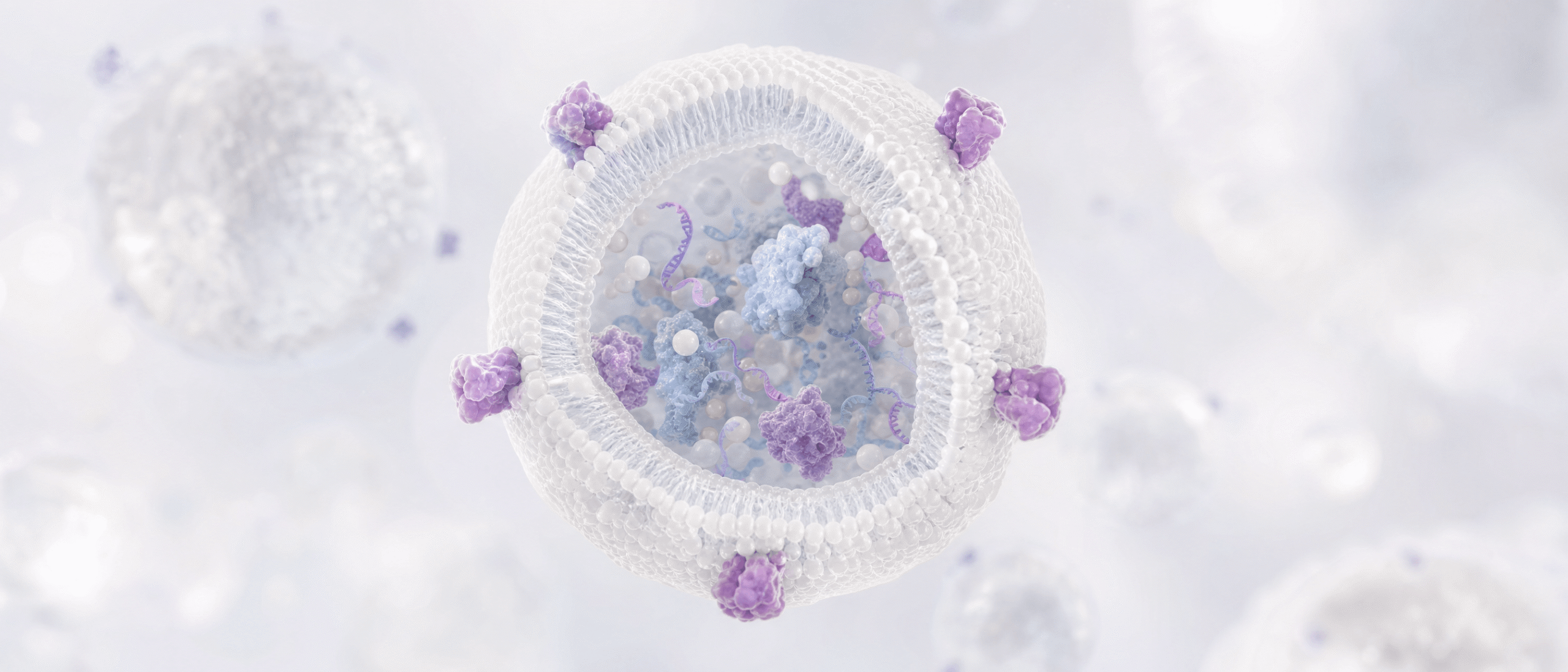
What Exosomes Are
The Producing Cell Matters
Exosomes used in research are most often collected from the culture medium of cells grown in the laboratory. The parent cell type shapes the vesicle cargo. Vesicles from umbilical cord MSCs are not the same as vesicles from adipose-derived cells, and both differ from vesicles taken from blood plasma.
Culture conditions also matter:
- Serum-free or vesicle-depleted media prevent animal or human serum vesicles from mixing into the preparation.
- Cell passage number can change what the cells secrete.
- Oxygen levels and cell density can alter vesicle output and content.
Exosome Isolation Methods Compared
Differential Ultracentrifugation
- Spins culture fluid at increasing speeds to remove cells, debris, and larger vesicles, then pellets the small vesicles
- Widely used and well documented [7]
- May co-isolate protein clumps and other particles, and very high forces can damage vesicles or cause them to aggregate
Density Gradient Ultracentrifugation
- Layers the sample in a density medium so vesicles settle at a characteristic density
- Often gives a purer preparation than a simple pellet
- Takes more time and yields less material
Size-Exclusion Chromatography (SEC)
- Passes the sample through a column that separates particles by size
- Tends to preserve vesicle structure and removes many free proteins
- Dilutes the sample, so a concentration step usually follows
Tangential Flow Filtration (TFF)
- Moves fluid across a membrane in a continuous loop, which separates and concentrates particles by size
- Scales more easily than ultracentrifugation, so it suits larger production runs
- Needs careful control of membrane type, pressure, and flow rate
Precipitation Kits and Immunoaffinity Capture
- Polymer precipitation is simple and fast, but it can pull down non-vesicle contaminants along with the vesicles.
- Immunoaffinity capture uses antibodies against vesicle surface proteins. It is selective, but it captures only vesicles carrying the chosen marker and can be costly at scale.
Many laboratories combine two methods, for example TFF followed by SEC, to improve purity. Comparing a single-step and a multi-step process is one of the most informative questions you can ask.
What the Method Tells You About a Product
Questions Worth Asking
- Which cells produced the vesicles, and at what passage?
- Was the culture medium serum-free or vesicle-depleted?
- Which isolation method or combination of methods was used?
- How were particle concentration and size measured?
- Were protein and non-vesicle contaminants measured?
- Was the final product filtered, lyophilized, or cryopreserved, and how was stability handled?
Signs of a Transparent Supplier
- A named isolation method, not a vague phrase like “proprietary process”
- Particle counts reported with the measurement technique, such as nanoparticle tracking analysis
- Test results for sterility, endotoxin, and mycoplasma
- Batch-level documentation that you can match to the lot in your hands
Be Careful With Particle Counts
How Exosomes Are Characterized
The ISEV guidelines, known as MISEV, recommend using several complementary tests rather than one [5][9]. A well-characterized preparation typically includes:
- Particle size and concentration: nanoparticle tracking analysis or a similar method
- Protein markers: presence of vesicle-associated proteins such as tetraspanins (CD9, CD63, CD81), plus absence of markers from other cell compartments
- Morphology: electron microscopy to look at the shape and structure of the particles
- Purity indicators: protein-to-particle ratio and tests for non-vesicle contaminants
- Safety testing: sterility, endotoxin, and mycoplasma
Quality Systems and Documentation
Good science depends on good records. Several quality frameworks apply to this field:
- cGMP (current Good Manufacturing Practice): written procedures for manufacturing, cleaning, and record keeping
- ISO 7 cleanroom classification: controls the number of airborne particles in the production space
- ISO 20387: a standard for biobanking that covers competence, impartiality, and traceability
- USP sterility and endotoxin methods: standard tests for microbial contamination and bacterial endotoxin
- Certificate of Analysis (COA): a lot-specific report listing the tests run and their results
A COA is only useful when it matches the lot you received. Check that the batch number on the document matches the vial, and read the actual results, not just the pass/fail summary.
Sourcing and Quality at CellGenic
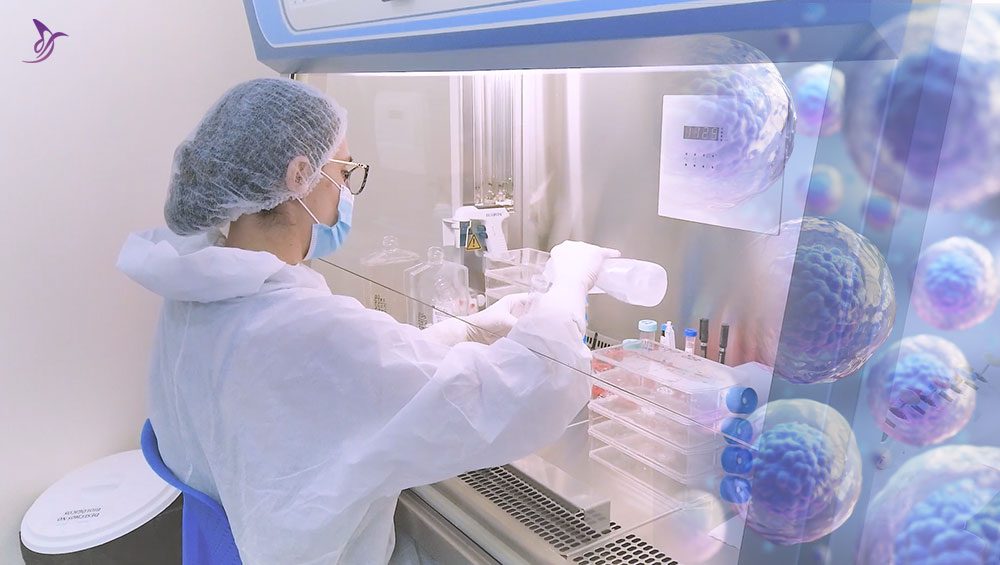
CellGenic is a provider-only supplier of research-grade cells, exosomes, and peptides. According to the company, products are made in its own cGMP-certified facility with an ISO 7 cleanroom, and its biobanking operations follow ISO 20387.
The company describes a five-stage chain of custody covering sourcing, laboratory work, QC and compliance, packaging, and shipping. Its stated approach includes:
- Sourcing: donor screening, raw-material qualification, and a certificate of origin for inputs
- Laboratory: cell expansion and vesicle isolation under one roof
- Testing: sterility, endotoxin, mycoplasma, identity, and potency checks, with a COA shipped with each batch
- Packaging and shipping: lyophilized or cryopreserved formats, tamper-evident packaging, and a temperature logger in each box
You can browse the catalog, which includes Wharton’s jelly MSCs and MUSE cells and exosome products such as ExoElite 70 and lyophilized exosomes. Qualified physicians, clinic owners, and researchers can apply for provider access to view pricing and place orders. CellGenic’s own authenticity verification page lets you check a product against its batch record.
CellGenic products are intended for verified professionals, and the company’s catalog labels peptides as for research use only. Anyone considering a biological product should discuss it with a qualified healthcare professional.
What Current Research Says
- Studies in cell culture and animal models have investigated how MSCs and their extracellular vesicles influence inflammation, tissue repair signals, and cell communication.
- Results vary between models, sources, and methods, which is one reason standardized reporting matters.
- Human clinical evidence is limited for most applications, and many trials are small or early-stage.
- Researchers continue to work on standardizing isolation and potency measurement so that results from different laboratories can be compared.
Potential Risks and Known Limitations
- Contamination risk: Biological products can carry microbial or endotoxin contamination if they are not made and tested properly.
- Immune reactions: Donor-derived material may provoke immune responses in some people.
- Batch variability: Biological materials vary between lots, even under controlled conditions.
- Method limits: No current method isolates only one vesicle subtype with complete purity.
- Unclear mechanisms: The mechanisms behind reported effects are not fully understood.
- Limited long-term data: Long-term safety information in humans is scarce for many of these products.
Regulatory Status
When to Consult a Healthcare Professional
Speak with a licensed healthcare professional before considering any cell- or exosome-based product, especially if you:
- Have a medical condition or take prescription medication
- Are pregnant or breastfeeding
- Have a history of immune or autoimmune problems
- Are unsure whether a product is lawful or appropriate in your region
Do not use research-grade materials as a substitute for medical care.
Key Takeaways
- Tissue source, donor screening, and culture conditions shape what a cell or exosome product contains.
- Isolation methods differ in purity, yield, and scalability, and many labs combine two methods.
- Particle counts alone can mislead, so look for multiple characterization methods.
- Batch-level documentation, including a COA that matches the lot, is a practical sign of transparency.
- Human clinical evidence remains limited, and regulatory rules vary, so professional guidance matters.
Frequently Asked Questions
References
- Wang HS, Hung SC, Peng ST, et al. Mesenchymal stem cells in the Wharton’s jelly of the human umbilical cord. Stem Cells. 2004;22(7):1330-1337.
- Kuroda Y, Kitada M, Wakao S, et al. Unique multipotent cells in adult human mesenchymal cell populations. Proc Natl Acad Sci U S A. 2010;107(19):8639-8643.
- Dominici M, Le Blanc K, Mueller I, et al. Minimal criteria for defining multipotent mesenchymal stromal cells. The International Society for Cellular Therapy position statement. Cytotherapy. 2006;8(4):315-317.
- Kalluri R, LeBleu VS. The biology, function, and biomedical applications of exosomes. Science. 2020;367(6478):eaau6977.
- Théry C, Witwer KW, Aikawa E, et al. Minimal information for studies of extracellular vesicles 2018 (MISEV2018): a position statement of the International Society for Extracellular Vesicles. J Extracell Vesicles. 2018;7(1):1535750.
- Gardiner C, Di Vizio D, Sahoo S, et al. Techniques used for the isolation and characterization of extracellular vesicles: results of a worldwide survey. J Extracell Vesicles. 2016;5:32945.
- Théry C, Amigorena S, Raposo G, Clayton A. Isolation and characterization of exosomes from cell culture supernatants and biological fluids. Curr Protoc Cell Biol. 2006;Chapter 3:Unit 3.22.
- Webber J, Clayton A. How pure are your vesicles? J Extracell Vesicles. 2013;2:19861.
- Welsh JA, Goberdhan DCI, O’Driscoll L, et al. Minimal information for studies of extracellular vesicles (MISEV2023): from basic to advanced approaches. J Extracell Vesicles. 2024;13(2):e12404.
- U.S. Food and Drug Administration. Public Safety Notification on Exosome Products. December 2019. https://www.fda.gov
- CellGenic. Manufacturing and quality information. https://cellgenic.com