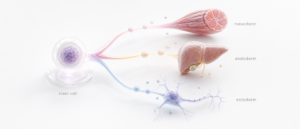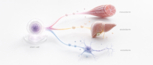
Exosomes are a specific subtype of extracellular vesicle: nanoscale, lipid-bilayer-bound particles that cells release into their surroundings, distinguished from other extracellular vesicles by where inside the cell they form. That detail carries the whole definition. [1][5][6]
Ask ten suppliers what an exosome is, and most will describe something close to a very small particle carrying signaling molecules. True enough, but also true of several other kinds of extracellular vesicle that aren’t exosomes at all. The International Society for Extracellular Vesicles’ current nomenclature standard, MISEV2023, is explicit on this point: “exosome” is a claim about biogenesis, specifically that a vesicle formed inside an endosomal compartment and was released when that compartment fused with the cell’s outer membrane. Unless that origin has actually been demonstrated for the material in question, the correct term is “small extracellular vesicle,” not “exosome.” [1][2]
That distinction is a reasonable proxy for how carefully a supplier actually understands what it’s selling. This guide covers what exosomes are, how they form, what they carry, how a lab confirms what’s actually in a vial, and why sourcing changes the answer.
The Word “Exosome” Is a Claim About Origin, Not Just Size
Extracellular vesicles, EVs, are the umbrella category: particles released from cells, enclosed by a lipid bilayer, incapable of replicating on their own. Within that umbrella sits a range of subtypes that differ by where and how they form inside the cell. Historically, the field split these roughly into two biogenesis-based groups: vesicles that bud directly from the plasma membrane, called ectosomes or microvesicles, and vesicles that form inside late endosomal compartments called multivesicular bodies, then get released when those compartments fuse with the plasma membrane. The second group is what “exosome” originally meant. [1][6][10]
The problem MISEV2023 addresses directly is that no isolation method available today can select for endosomal origin specifically. Ultracentrifugation, size-exclusion chromatography, precipitation, and every other common separation technique all pull down a mixed population of vesicles from multiple origins at once, because there’s no marker unique to the endosomal pathway. So when a lab or supplier isolates vesicles from conditioned media or a cell source and calls the output “exosomes,” it’s usually making a claim about origin the isolation method itself can’t actually support (Welsh et al., Journal of Extracellular Vesicles, 2024).
MISEV2023 answers this with operational language wherever demonstrated origin isn’t available: “small EV” for isolated vesicles generally under 200 nanometers in diameter, and “large EV” for those above that threshold. Biogenesis terms like “exosome” and “ectosome” are reserved for cases where subcellular origin has actually been shown [1][2] for that specific source and condition. In most preparations on the market, that’s the exception, not the rule.
This is the single most useful filter a clinician has for evaluating a supplier’s technical literature. A company that consistently and correctly uses “small extracellular vesicle,” except where it has evidence for endosomal origin, is describing its product accurately. A company whose every page says “exosome” without ever touching this distinction is, at best, using industry shorthand loosely, and at worst, making an origin claim it hasn’t verified.
Extracellular Vesicle Subtypes at a Glance
A quick reference, since these terms get used inconsistently across the literature and across supplier marketing.
Extracellular vesicle (EV): The umbrella term for any particle released from a cell that is enclosed by a lipid bilayer and cannot replicate on its own. Covers exosomes, ectosomes, apoptotic bodies, and other membrane-bound vesicles of cellular origin. [1][4][10]
Exosome: A biogenesis-specific subtype of small EV that forms as an intraluminal vesicle inside a multivesicular body and is released when that compartment fuses with the plasma membrane. The term is accurate only when endosomal origin has been demonstrated for the specific preparation.
Small extracellular vesicle (sEV): The MISEV2023-recommended operational term for isolated vesicles generally under 200 nanometers in diameter, used when biogenesis hasn’t been confirmed. This is the accurate default term for most commercially isolated preparations.
Ectosome (microvesicle): A vesicle that buds directly outward from the plasma membrane rather than forming inside an internal compartment first. Ectosomes overlap in size with small EVs, which is part of why size alone can’t distinguish the two.
Apoptotic body: A larger vesicle released during programmed cell death, generally well outside the small-EV size range, structurally and functionally distinct from exosomes and ectosomes.[1][10]
Sizes commonly cited for exosomes specifically, roughly 30 to 150 nanometers, come from earlier literature that predates the field’s current operational framework. That range was never tied to a validated origin-specific marker. MISEV2023’s own threshold for the broader small-EV category sits under 200 nanometers, and that figure carries current consensus behind it.
How Exosomes Form: The Endosomal Pathway
The biogenesis pathway that defines a true exosome starts inside the cell, not at the membrane. It begins when a segment of the endosomal membrane buds inward, into the lumen of the endosome itself, rather than outward into the extracellular space. Repeated inward budding produces a compartment called a multivesicular body, packed with these intraluminal vesicles. From there, one of two things happens: the multivesicular body can fuse with a lysosome, in which case its contents are degraded, or it can traffic to and fuse with the plasma membrane, releasing its intraluminal vesicles into the extracellular environment. Those released vesicles are exosomes.[1][6][8][10]
An ectosome forms differently. It buds directly outward from the plasma membrane in a single step, never passing through an internal endosomal compartment. The two pathways involve different membrane trafficking machinery and different sorting of cargo into the forming vesicle. That’s part of why exosomes and ectosomes can carry meaningfully different molecular content even when they end up similar in size (Zhang et al., Cell & Bioscience, 2019). [6][8]
None of this happens in isolation from the cell’s broader physiology. Which cargo gets sorted into forming intraluminal vesicles, how actively a given cell type produces them, and under what conditions release increases are active areas of ongoing research, not settled points. What’s well established is the structural mechanism: endosomal origin, intraluminal vesicle formation, multivesicular body trafficking, and plasma membrane fusion as the release step.
What’s Inside: Cargo and Function
Exosomes and other small EVs carry a mixed cargo of proteins, lipids, and nucleic acids, including microRNA and messenger RNA fragments, enclosed and protected by the lipid bilayer that forms the vesicle’s outer boundary. That enclosure matters functionally. It’s what allows this cargo to travel through extracellular fluid without being degraded the way free-floating RNA or protein would be almost immediately.
The generally accepted mechanism of action is paracrine signaling: a released vesicle is taken up by, or fuses with, a nearby or distant target cell, delivering its cargo and potentially influencing that cell’s gene expression or signaling state. Cell-based approaches like mesenchymal stem cell administration work differently. A living cell is introduced, and its functional role depends partly on that cell’s own ongoing activity and migration. A small EV carries a molecular payload and nothing more. It isn’t alive, and it can’t divide or engraft.
Surface composition matters as much as internal cargo. Small EVs typically display a set of membrane proteins, including tetraspanins such as CD9, CD63, and CD81, that are commonly used across the field as positive identity markers, though none of them is exclusively specific to endosomal-origin vesicles on its own. That limitation is exactly why MISEV2023 calls for a multi-marker, multi-method characterization approach rather than reliance on any single marker.
How a Lab Actually Confirms What’s in a Vial
Because no single marker is exosome-specific, MISEV2023 recommends characterizing a preparation across several independent categories: [1][2]
- Particle concentration and size distribution, typically measured by nanoparticle tracking analysis or a comparable method, to confirm the preparation actually falls in the expected size range rather than being dominated by larger contaminants or protein aggregates.
- Positive protein markers, generally including at least one tetraspanin (CD9, CD63, or CD81) and at least one cytosolic protein associated with EV biogenesis, confirmed by a method such as Western blot or flow cytometry.
- Negative markers, proteins that should be absent or minimal in a clean preparation, used to flag contamination from other cellular compartments or co-isolated debris.
- Morphological confirmation, typically by electron microscopy, since size and marker data alone don’t confirm that the isolated particles are actually intact, single-membrane vesicles rather than aggregates or artifacts.
Each category checks something the others can’t, which is why labs run them together rather than leaning on any single result. [1][4]
[VERIFY: the exact marker panel CellGenic’s lab runs per batch, including which positive and negative markers are tested and by which method. Placeholder until confirmed; do not publish with an invented marker list.]
This is where a supplier’s documentation either holds up or doesn’t. A Certificate of Analysis that lists a specific, named marker panel tied to a specific batch number, alongside particle-size data and a stated isolation method, describes an actual characterized preparation. A COA that asserts purity or identity without naming what was tested is asserting a conclusion without showing the work behind it. How that isolation happens in the first place, and what it does and doesn’t select for, is its own question, covered in our guide to how exosomes are isolated and obtained.
Sourcing Determines What You’re Actually Buying
Cell or tissue source shapes exosome composition as much as biogenesis pathway does. Vesicles isolated from mesenchymal stem cells, induced pluripotent stem cells, and plant tissue are not interchangeable starting materials, even when each gets marketed under the same general “exosome” label. They differ in the cell types that produced them, the cargo those cell types tend to package, and in some cases, how carefully the biogenesis pathway itself has been characterized for that source. MSC-derived material varies further by tissue of origin. Bone marrow, adipose tissue, and umbilical cord sources produce MSCs with measurably different secreted profiles. That’s a sourcing detail suppliers rarely spell out on their own.
Our [comparison of MSC, iPSC, and plant-derived exosome sourcing](URL-TBD: msc-ipsc-plant-exosome-sourcing) covers how these starting materials differ in practice. For CellGenic’s cell-therapy line, which shares some sourcing considerations but is a structurally distinct product category from exosomes, see the [Muse cell pillar page](URL-TBD: muse-cell-pillar-page).
Why This Distinction Matters for a Research Protocol
Getting exosome nomenclature right functions as a proxy for something bigger: whether a supplier’s other technical claims, about sterility, potency, and batch consistency, get handled with the same precision or the same looseness. A company that can tell you exactly what biogenesis claim it is and isn’t making about its own material is a company whose Certificate of Analysis is probably worth trusting on everything else.
If you’re weighing exosome-derived material against a cell-based approach for a given protocol, our comparison of exosomes and mesenchymal stem cells covers how the two actually differ, beyond terminology. Before introducing any extracellular vesicle preparation into a research protocol, our [overview of exosome safety considerations](URL-TBD: exosome-safety-considerations) is worth reading alongside this guide.
Providers evaluating CellGenic as a source for research-use extracellular vesicle products can apply for provider access to review batch-specific documentation directly.
Common Questions
Is “exosome” just a marketing term, or does it mean something specific?
Per MISEV2023, “exosome” refers to a vesicle of demonstrated endosomal origin, a mechanistic claim that most marketing copy quietly drops. If that origin hasn’t been shown for a given preparation, “small extracellular vesicle” is the accurate term to use instead.
Why does endosomal origin matter if the vesicles look and behave similarly either way?
Origin affects cargo sorting, surface composition, and the biogenesis machinery involved, all of which can meaningfully affect what a preparation actually contains, even between vesicles of similar size. Treating “exosome” as a size category rather than an origin category obscures a real biological distinction.
Can a supplier legitimately call its product “exosomes” without confirming endosomal origin?
Only if that origin has actually been demonstrated for the specific source and condition, which most commercial isolation methods can’t establish on their own. [1][7] In practice, most preparations on the market are more accurately described as small extracellular vesicles than as exosomes in the strict MISEV2023 sense.
What markers should I expect a supplier to test for, and why isn’t one marker enough?
No single marker is unique to endosomal-origin vesicles. That’s why MISEV2023 recommends a multi-marker, multi-method approach: positive tetraspanin markers, negative contamination markers, particle-size data, and morphological confirmation, checked together rather than in isolation.
How does exosome sourcing (MSC, iPSC, plant-derived) affect what’s in the vial?
The producing cell or tissue type shapes cargo composition and, in some cases, how well the biogenesis pathway itself has been characterized. Material from different sources isn’t interchangeable, even when labeled with the same general term.
Does a correctly labeled “small extracellular vesicle” product mean it’s lower quality than one labeled “exosome”?
No. A correctly labeled small extracellular vesicle product is simply describing its demonstrated origin accurately, rather than making an unverified biogenesis claim. Precision in labeling signals a supplier who understands what they’re actually selling.
References
- Welsh JA, Goberdhan DCI, O’Driscoll L, et al. Minimal Information for Studies of Extracellular Vesicles 2023 (MISEV2023): From Basic to Advanced Approaches. Journal of Extracellular Vesicles. 2024;13(2):e12404. doi:10.1002/jev2.12404. Full textpmc.ncbi.nlm.nih
- International Society for Extracellular Vesicles. MISEV2023: Minimal Information for Studies of Extracellular Vesicles. Available at: ISEV.isev
- Welsh JA, Goberdhan DCI, O’Driscoll L, et al. Minimal Information for Studies of Extracellular Vesicles 2023: From Basic to Advanced Approaches. Journal of Extracellular Vesicles. 2024;13(2):e12404. Publisher and repository record.eprints.whiterose.ac
- Théry C, Witwer KW, Aikawa E, et al. Minimal Information for Studies of Extracellular Vesicles 2018 (MISEV2018): Position Statement of the International Society for Extracellular Vesicles and Update of MISEV2014. Journal of Extracellular Vesicles. 2018;7(1):1535750. doi:10.1080/20013078.2018.1535750. PMC
- Kalluri R, LeBleu VS. The Biology, Function, and Biomedical Applications of Exosomes. Science. 2020;367(6478):eaau6977. doi:10.1126/science.aau6977.
- van Niel G, D’Angelo G, Raposo G. Shedding Light on the Cell Biology of Extracellular Vesicles. Nature Reviews Molecular Cell Biology. 2018;19:213–228. doi:10.1038/nrm.2017.125.
- Doyle LM, Wang MZ. Overview of Extracellular Vesicles, Their Origin, Composition, Purpose, and Methods for Exosome Isolation and Analysis. Cells. 2019;8(7):727. doi:10.3390/cells8070727. PMCpmc.ncbi.nlm.nih
- Zhang Y, Liu Q, Zhang X, Huang H, Tang S, Chai Y, Xu Z, Li M, Chen X. Exosomes: Biogenesis, Biologic Function and Clinical Potential. Cell & Bioscience. 2019;9:19. doi:10.1186/s13578-019-0282-2. PubMedpmc.ncbi.nlm.nih
- Mathieu M, Martin-Jaular L, Lavieu G, Théry C. Specificities of Secretion and Uptake of Exosomes and Other Extracellular Vesicles for Cell-to-Cell Communication. Nature Cell Biology. 2019;21:9–17. doi:10.1038/s41556-018-0250-9.
- Raposo G, Stoorvogel W. Extracellular Vesicles: Exosomes, Microvesicles, and Friends. Journal of Cell Biology. 2013;200(4):373–383. doi:10.1083/jcb.201211138.
- Théry C, Ostrowski M, Segura E. Membrane Vesicles as Conveyors of Immune Responses. Nature Reviews Immunology. 2009;9:581–593. doi:10.1038/nri2567.
REGULATORY DISCLAIMER · EXOSOMES These products are intended for laboratory research use only. They are not drugs, foods, cosmetics, or medical treatments and must not be used for any form of human or animal administration. All information provided is for educational and scientific reference only and the products should be handled exclusively by licensed, qualified professionals. Misbranding, misuse, or mislabeling of these products as therapeutic or consumable substances is strictly prohibited by law.