The claim that a cell administered intravenously finds an injury on its own is the part of the Muse literature that sounds least plausible and is best evidenced. It also gets described more often than it gets explained.
What follows is the mechanism as published: how the cells locate damage, what happens when they arrive, what changes in the tissue afterwards, and, because this is where most writing on the subject fails, what the animal evidence does not extend to.
Three steps, each separately evidenced
Three steps, each separately evidenced in preclinical models.
The cells sense a signal released by damaged tissue and accumulate there after intravenous administration rather than distributing evenly. Having arrived, they engraft and differentiate into the cell types of the host tissue without induction. And the tissue environment changes measurably around them, with less fibrosis and less inflammation. [1][2][3][4]
Every study below is an animal study. Nothing on this page describes a result in humans; for that, see where the clinical research currently stands. For what the cells are before any of this begins, see what Muse cells are and how they were identified.
Step1: How the cells find the injury
The S1P–S1PR2 axis
The signal is sphingosine-1-phosphate, a lipid mediator released by damaged tissue, and the receptor is sphingosine-1-phosphate receptor 2. [4]
Yamada and colleagues established this in Circulation Research in 2018, working in a model of acute myocardial infarction. Muse cells express S1PR2 and migrate along S1P gradients emanating from injured tissue, which is what makes the distribution selective rather than systemic; the cells accumulate where the signal originates. The same work reported that donor cells survived in host tissue as differentiated cells for more than 6 months without immunosuppression.
That is the reason Muse cells are home to damaged tissue rather than lodging wherever the circulation happens to take them. It is a receptor-ligand mechanism, not a property. [4]
CXCR4, and an incomplete answer
The most quantitatively detailed evidence of homing comes from a different organ. Iseki and colleagues, publishing in Cell Transplantation in 2017, used a carbon tetrachloride model of liver fibrosis in immunodeficient mice, administering 5 × 10⁴ human Muse cells by tail vein at weeks two, four, and six. [2]
They then measured where the cells went, using quantitative PCR for human-specific Alu sequences. Muse cells showed their highest Alu signal in the liver at two weeks. Non-Muse cells from the same source showed no detectable liver signal at all. The same starting material, minus the SSEA-3-positive fraction, did not home.
That study also probed the mechanism and reported an incomplete answer, which is worth repeating precisely because it is a limitation. Blocking CXCR4 with the antagonist AMD3100 substantially suppressed Muse cell migration toward damaged serum, but did not abolish it. The authors concluded that additional, unidentified factors contribute. So, the fact that Muse cells are home to injury sites is well established across at least two organs and two mechanisms, and the full account of how is not finished. [2][4]
Step 2: engraftment and spontaneous differentiation
Arrival is not the interesting part. What the cells do next is
In the liver fibrosis model above, integrated Muse cells differentiated into functional liver cells with no pre-transplantation induction. Of the integrated cells, 71.1 ± 15.2% expressed HepPar-1, 54.3 ± 8.2% expressed human albumin, and 47.9 ± 4.6% expressed anti-trypsin. Roughly 97% did so without cell fusion with host hepatocytes, meaning the human cells became hepatocyte-like cells themselves rather than merging with existing ones.
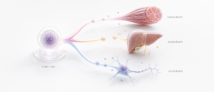
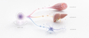
That is spontaneous, tissue-appropriate differentiation directed by the environment the cells arrived in. It is the mechanistic reason the same population has been studied across unrelated organs, and it depends on the triploblastic capacity described in how the Muse fraction sits inside a mesenchymal preparation.
Step three: fibrosis and inflammation
Two measurable changes follow, and they are the outcomes most often reported.
That Muse cells reduce fibrosis has been quantified. In the liver model, Sirius red staining put the fibrotic area at 0.75 ± 0.15% in treated animals against 2.91 ± 0.35% in vehicle controls. The authors attributed part of that effect to Muse cell production of matrix metalloproteinase-9. In the hindlimb ischemia model discussed below, the fibrotic area also decreased significantly compared to controls.
Anti-inflammatory effects and the mediators behind them
The anti-inflammatory effects are attributed to named mediators rather than to a general immunomodulatory reputation. Kuroda and colleagues, reviewing the mechanism in Frontiers in Pharmacology in 2022, report that Muse cells produce the anti-inflammatory cytokines IL-10 and TGF-β at levels above non-Muse mesenchymal cells, along with EGF, KGF, VEGFA and PDGF variants, and that macrophages significantly reduce TNF-α production when co-cultured with Muse cells in vitro.[3]
The immune-privilege side is the same review’s other contribution: approximately 90% of Muse cells express HLA-G, compared with roughly 5% of mesenchymal stromal cells, alongside production of IDO, TGF-β, PGE2, and HGF, which act on regulatory T-cell maturation and lymphocyte proliferation. The relevance to the mechanism is that cells that persist in tissue can exert an anti-inflammatory and anti-fibrotic effect over time rather than transiently. The immunological detail belongs with the safety literature: what the published safety literature reports.
Limb regeneration: no study reports regrowth
This needs addressing directly, because Muse stem cells limb regeneration is one of the most searched phrases in this subject, and it does not describe anything in the literature.[1]
What exists and what searches for Muse cells for limb regeneration are almost certainly reaching for a single study of hindlimb ischemia. Hori and colleagues, in Frontiers in Cardiovascular Medicine in 2022, induced ischemia in mice by ligating the femoral artery and vein, then administered 3 × 10⁴ human Muse cells intravenously one day later, with no immunosuppressant, against non-Muse mesenchymal cells and saline controls.
At postoperative days 7 and 14, laser Doppler measurement showed significantly higher blood flow in the Muse group than in controls. Microvascular density rose, measured as CD31-positive cells; fibrotic area fell; and labelled Muse cells were found in ischemic border zones expressing the vascular endothelial marker CD31.
Recovery of blood flow to an ischemic limb is a meaningful finding. It is not limb regeneration, and no limb or tissue regrowth was reported in that study. The distance between those two statements is roughly the distance between this page and the clinic pages that rank alongside it.
Four limits on the regeneration evidence
Muse stem cells’ regeneration potential is real, specific, and bounded. Anyone assessing Muse stem cells for tissue regeneration should hold the evidence and the limits together.
The evidence: selective homing by a named receptor mechanism, engraftment without induction, tissue-appropriate differentiation at measurable rates, quantified reduction in fibrotic area, and named anti-inflammatory mediators reproduced across unrelated organ systems.
The limits, in order of how often they are ignored. These are animal models in mice and rats, several of them immunodeficient. The doses are small, and the observation windows are weeks to months, not years. Cell numbers, routes, and timings differ between studies, so results are not directly comparable. And a mechanism demonstrated in a model animal is a hypothesis about humans, not a finding in them. [1][2][3][4]
Where do Muse cells sit in regenerative medicine?
Interest in Muse cells in regenerative medicine follows from the mechanism rather than from the outcomes. A population that homes selectively, engrafts without induction, and differentiates according to its destination is doing something structurally different from a cell administered for a paracrine effect alone. [2][4]
That is why Muse cells’ regenerative medicine research has spread across cardiac, hepatic, neurological, and vascular models rather than concentrating in one. And it is also why the research remains early: a mechanism that generalises still has to be demonstrated indication by indication, in humans, with controls.
Nothing on this page establishes that Muse cells treat, cure, prevent or diagnose any condition, and nothing on it supports administration outside a properly authorised research setting under applicable regulation.
At CellGenic, certificates of analysis are issued per batch and released to verified accounts. Access begins with provider verification.
Common questions
Can Muse cells regenerate a limb?
No published study reports the regrowth of a limb, or of any substantial anatomical structure, in any species. The nearest real work is a mouse hindlimb ischemia study that reported recovered blood flow, increased microvascular density, and reduced fibrosis, not regrowth.
How do Muse cells know where the injury is?
Through a receptor-ligand mechanism rather than a property. Damaged tissue releases sphingosine-1-phosphate; Muse cells express sphingosine-1-phosphate receptor 2 and migrate along that gradient, which is why distribution after intravenous administration is selective rather than systemic. 4]
Do Muse cells reduce fibrosis?
In animal models, measurably. In a mouse liver fibrosis model, Sirius red staining showed a fibrotic area of 0.75 ± 0.15% in treated animals, compared with 2.91 ± 0.35% in vehicle controls, attributed partly to matrix metalloproteinase-9 production. [1]
What is Muse stem cell regeneration potential, in practice?
Real, specific, and bounded. Selective homing, engraftment without induction, tissue-appropriate differentiation, and reduced fibrosis have been reproduced across unrelated organ systems, all in animal models, at small doses, over weeks to months, with study designs that are not directly comparable to each other.
References
Sources cited or paraphrased in this article are listed alphabetically by first author.
- Hori Y, Kitani T, Yanishi K, et al. “Intravenous administration of human Muse cells recovers blood flow in a mouse model of hindlimb ischemia.” Frontiers in Cardiovascular Medicine. 2022;9:981088. PMID: 36440014
- Iseki M, Kushida Y, Wakao S, Akimoto T, Mizuma M, Motoi F, Asada R, Shimizu S, Unno M, Chazenbalk G, Dezawa M. “Human Muse cells, nontumorigenic pluripotent-like stem cells, have liver regeneration capacity through specific homing and cell replacement in a mouse model of liver fibrosis.” Cell Transplantation. 2017;26(5):821–840. DOI: 10.3727/096368916X693662
- Kuroda Y, Oguma Y, Hall K, Dezawa M. “Endogenous reparative pluripotent Muse cells with a unique immune privilege system: hint at a new strategy for controlling acute and chronic inflammation.” Frontiers in Pharmacology. 2022;13:1027961. DOI: 10.3389/fphar.2022.1027961
- Yamada Y, et al. “S1P–S1PR2 axis mediates homing of Muse cells into damaged heart for long-lasting tissue repair and functional recovery after acute myocardial infarction.” Circulation Research. 2018. PMID: 29475983
REGULATORY DISCLAIMER · CELLS
These products are intended for laboratory research use only. They are not drugs, foods, cosmetics, or medical treatments and must not be used for any form of human or animal administration. All information provided is for educational and scientific reference only and the products should be handled exclusively by licensed, qualified professionals. Misbranding, misuse, or mislabeling of these products as therapeutic or consumable substances is strictly prohibited by law.